

Cancer Research
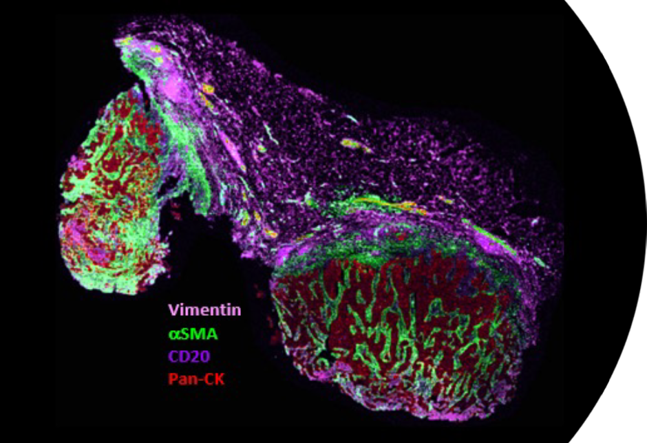
Miralys MALDI-IHC Imaging of a Breast Tumor Sample
Miralys imaging and its high-plex, multiomic and multimodal workflows can provide crucial new layers of information helping researchers achieve valuable insights for cancer research and shorten the transition to clinical applications. Examples include:
- Tumor Microenvironment: Cancer is a complex ecosystem comprising various heterogenous cell types, including immune cells, endothelial cells and fibroblasts, among others. Understanding the spatial organization and interaction of these components within the tumor microenvironment is essential for comprehending cancer progression, metastasis, and treatment responses.
- Spatial Heterogeneity: Tumors exhibit spatial heterogeneity, meaning different regions of the tumor may have distinct phenotypic characteristics. Spatial biology techniques allow researchers to map this heterogeneity, providing insights into the evolution of tumors, drug resistance mechanisms, and potential therapeutic approaches.
- Drug Distribution and Colocalization: Miralys imaging and associated work flows can be used to image the distribution of unlabeled small molecule drugs along with targeted macromolecules such as proteins in a tissue. This provides valuable information such as the effects of drugs on expression levels of downstream pathways, and colocalization of the drugs and target or untargeted receptors. Using this information, researchers can identify potential causes of toxicity and design more effective treatment strategies.
- Immune Response: Spatial biology allows for the characterization of spatial patterns of immune infiltration within tumors. Understanding the spatial distribution of immune cells and their interactions with cancer cells provides insights into the tumor immune landscape, which is critical for developing immunotherapies and predicting patient outcomes.
Miralys-powered spatial biology provides a deeper understanding of the spatial organization and dynamics of tumors, which is essential for unraveling the complexity of cancer biology and improving diagnostic and therapeutic strategies.
Miralys: A Versatile Tool for Studying Neurodegenerative Diseases
Fast, ultra-high plex Miralys imaging can help us better understand neurodegenerative diseases.
Spatial Mapping of Disease-State Models. Animal models, such as mice, rats, and non-human primates, can be genetically engineered or exposed to toxins to replicate key features of neurodegenerative diseases. These models often develop pathological hallmarks similar to those observed in humans, such as protein aggregation, neuronal loss, and behavioral deficits, allowing researchers to study disease progression and test potential interventions. Miralys has been used, for example, to study models such as these in Alzheimer’s and Parkinson’s disease.
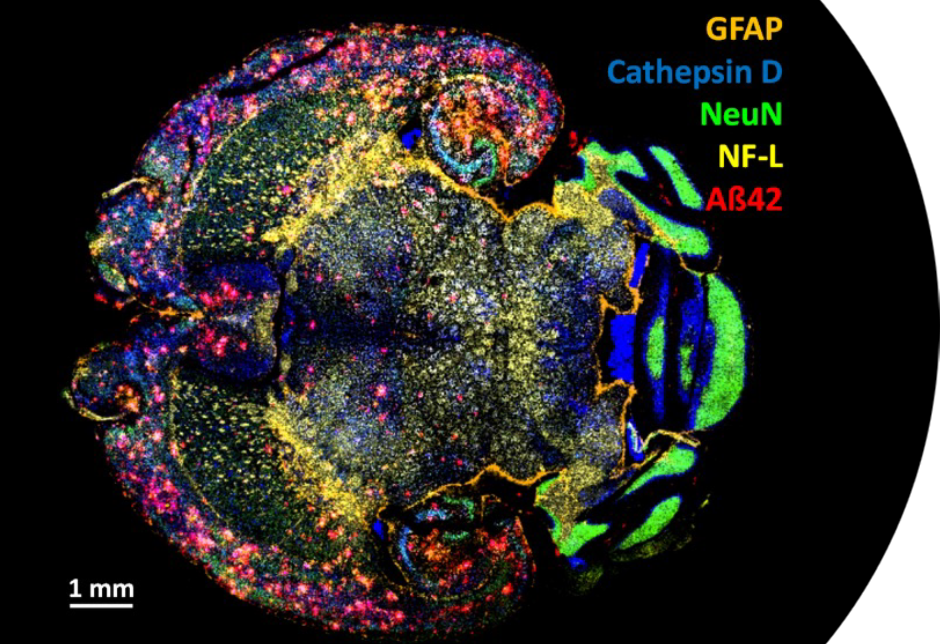
A Miralys MALDI-IHC image of a radial section of an Alzheimer’s model mouse brain. The pink flower-like clusters in the top and bottom left of this image are Amyloid plaques such as those that occur in human Alzheimer’s Disease.