Enabling Multiomics

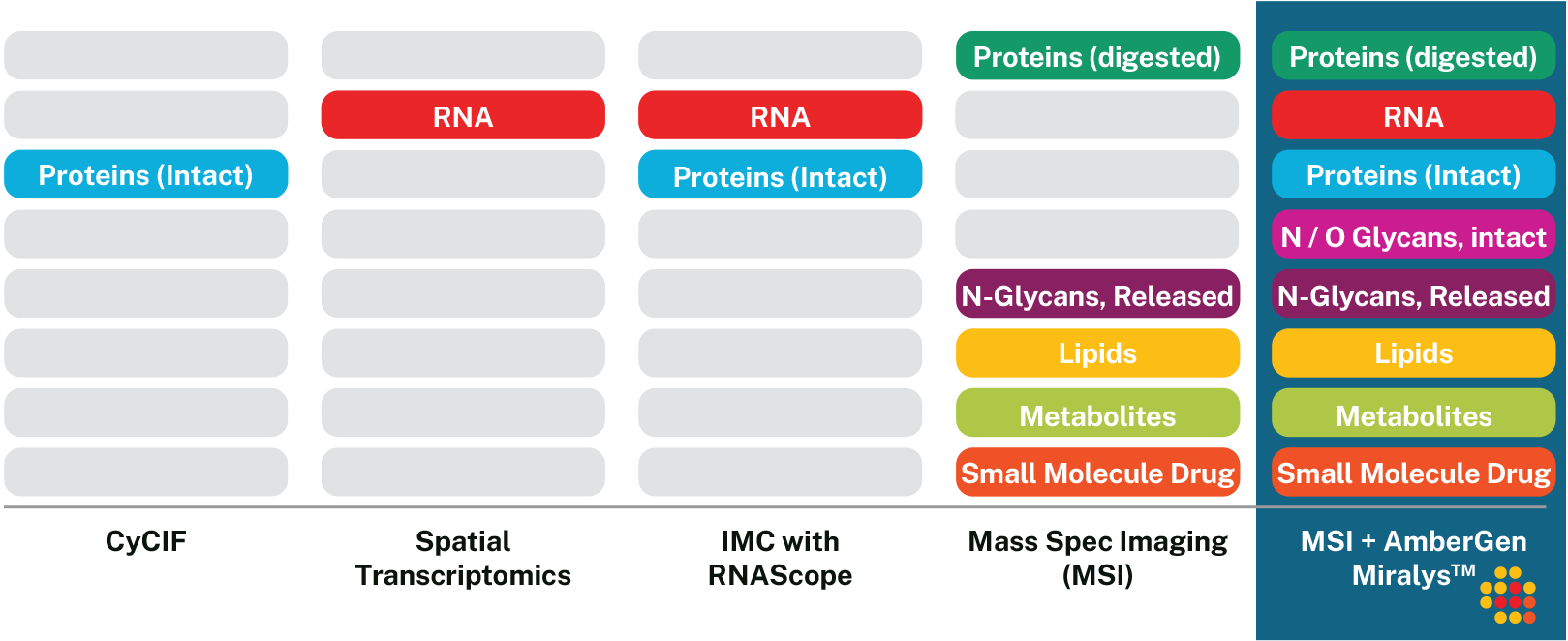
Miralys™ delivers the broadest spatial multiomic imaging in mass spectrometry

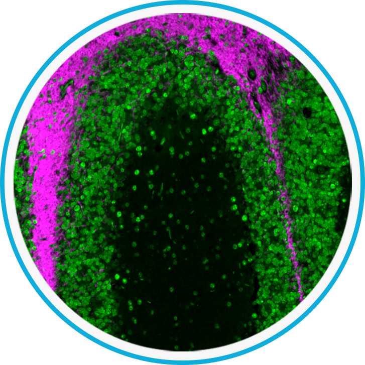
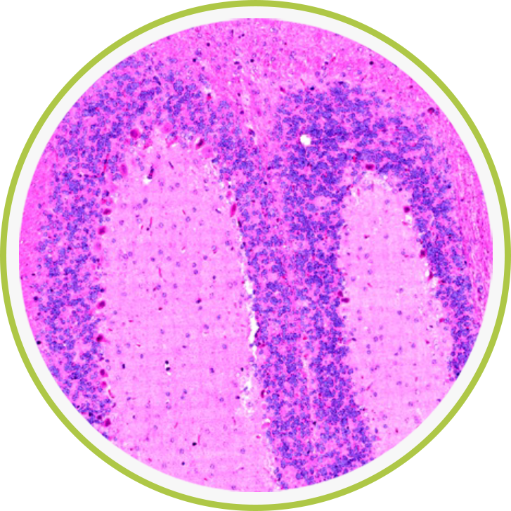
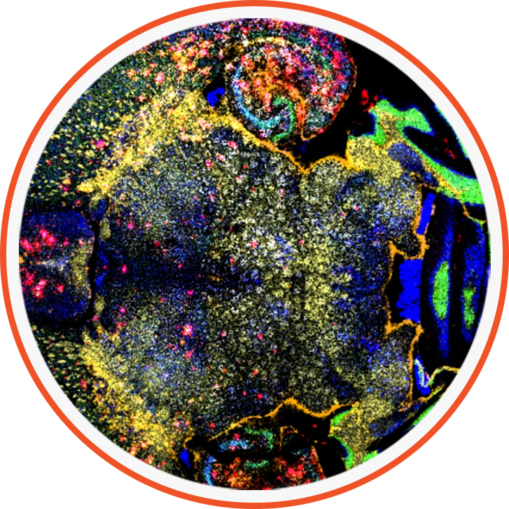
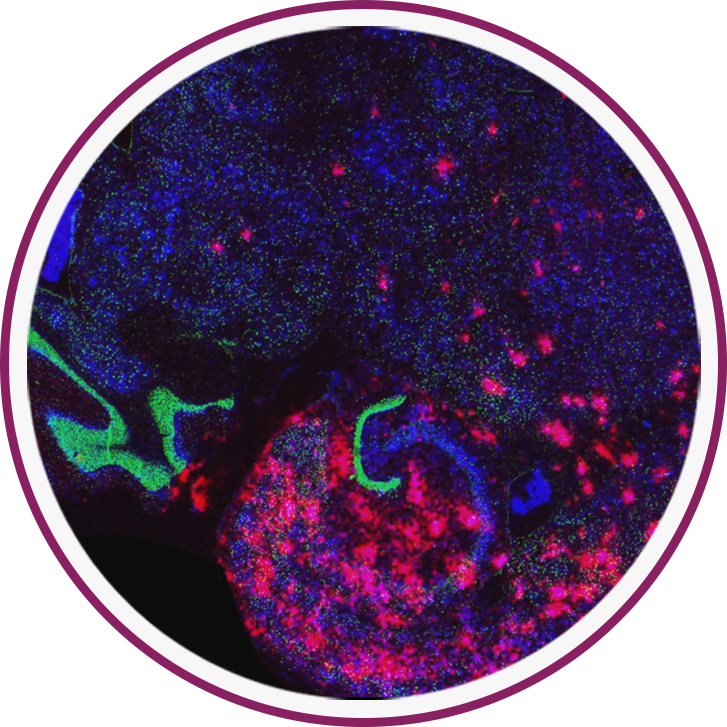
Spatial multiomic imaging reveals spatial patterns of of biomolecules within tissues, helping scientists understand complex biological systems, uncover disease mechanisms, guide precision medicine, accelerate drug discovery, and foster biomedical innovation.

Miralys builds upon the already strong spatial multiomic capabilities of matrix-assisted laser-desorption/ionization (MALDI) imaging to add new layers of biological insight. Now, researchers can visualize over 100 intact targeted protein distribution in the same tissue sample in which they have already imaged unlabeled small molecule such as drugs, lipids and other metabolites.

Miralys delivers the broadest single instrument imaging capability

Miralys extends the multiomic capabilities of MALDI imaging.
It lets you perform spatial multiomic imaging on the same sample with the same workflow, providing new insights.

Correlative Multimodal Imaging

Multimodal, Non-Destructive Imaging: Extract the Maximum Data from Each Tissue Sample

Correlative Multimodal Imaging integrates two or more imaging techniques to capture diverse details about a single sample. This approach generates a more comprehensive profile of the specimen, offering insights into its macroscopic, mesoscopic, and microscopic characteristics, including its structure, dynamics, function, and chemical makeup. Mass Spec Imaging and Miralys MALDI-IHC are especially well-suited for use in multimodal workflows. Various imaging modalities can be performed either before or after MALDI-IHC imaging.


The Most Multimodal

With Miralys MALDI-IHC, you can combine both small and large molecule imaging capabilities with other modalities such as fluorescent imaging, or laser capture microdissection followed by LC-MS, for example.
previous next
previous next
previous next
previous next

Miralys dual-labeled antibody probes which incorporate both mass-tags and fluorophores enable both immunofluorescence and MALDI-IHC imaging enabling accurate alignment of targeted proteins using both modalities*.

** M. J. Lim, G. Yagnik, C. Henkel, S. F. Frost, T. Bien and K. J. Rothschild (2023), MALDI HiPLEX-IHC: multiomic and multimodal imaging of targeted intact proteins in tissues In Front Chem 2023 Vol. 11 Pages 1182404 MALDI HiPLEX-IHC: multiomic and multimodal imaging of targeted intact proteins in tissues – PMC (nih.gov)

“…we have developed and demonstrated a workflow for using targeted MALDI-IHC imaging to guide an untargeted bottom-up spatial proteomics analysis with LC-MS revealing extensive intratumor heterogeneity of over-represented pathways. The complementarity of the two approaches is promising with regard to the further development of the spatial omics field.” *

* Claes, B. S. R., Krestensen, K. K., Yagnik, G., Grgic, A., Kuik, C., Lim, M. J., Rothschild, K. J., Vandenbosch, M., & Heeren, R. M. A. (2023). MALDI-IHC-Guided In-Depth Spatial Proteomics: Targeted and Untargeted MSI Combined. In Analytical Chemistry (Vol. 95, Issue 4, pp. 2329–2338). American Chemical Society (ACS). https://doi.org/10.1021/acs.analchem.2c04220

Hyperplex and Fast

Scan in Minutes, not Days

High-plex no longer means slow. With Miralys MALDI-IHC, a single scan can image more than 150 intact proteins in as fast as 45 minutes for a 1 cm2 tissue sample. Scan duration is independent of multiplexity, with no cycling required. Because it is not limited by broad fluorescence emission and excitation bands, Miralys is immune to spectral overlap and autofluorescence, allowing vastly more biomarkers to be imaged in a single scan.

When Miralys is used on industry-leading MALDI-MSI instrumentation, it can rapidly scan an entire 25 mm x 75 mm microscope slide containing multiple sample specimens. This capability makes Miralys ideal for analysis of tissue microarrays (TMAs) containing up to 100 separate tissue samples. The ability to perform successive Miralys™ MALDI-IHC imaging runs means that after a rapid scan one can zoom in at cellular resolution to regions of interest on a sample.

Hyperplex illustration

32 Human organ TMA imaged at 49-plex using MALDI-IHC. End-to-end imaging of this sample,
including sample preparation, was accomplished in 24 hours.

Miralys is so fast, many adjacent tissue slices from a block can be imaged in succession,
allowing reconstruction of 3D proteomic images.

Co-localize Small Molecule Drugs and their Targets

Use Miralys to add exciting new capabilities to Mass Spec Imaging

Mass Spec Imaging (MSI) has become ubiquitous in Pharmaceutical Development over the past decade due to its ability to map drug distribution / retention in tissue using label-free techniques. Now Miralys can be used to map proteins, in an ultra-high-plex manner, on the same tissue sample, and on the same instrument. The combined approach can help drug developers understand if drugs are reaching their targets.